MacDonald Environmental Services
Home
Gallery
Diagnostic Microscopy Services
Training Services
Contact
Check out these images from some wastewater treatment plants. Hover over the images to see captions. Use these images however you would like - but please credit Jeff MacDonald of MacDonald Environmental Services - thanks!
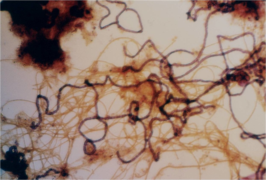
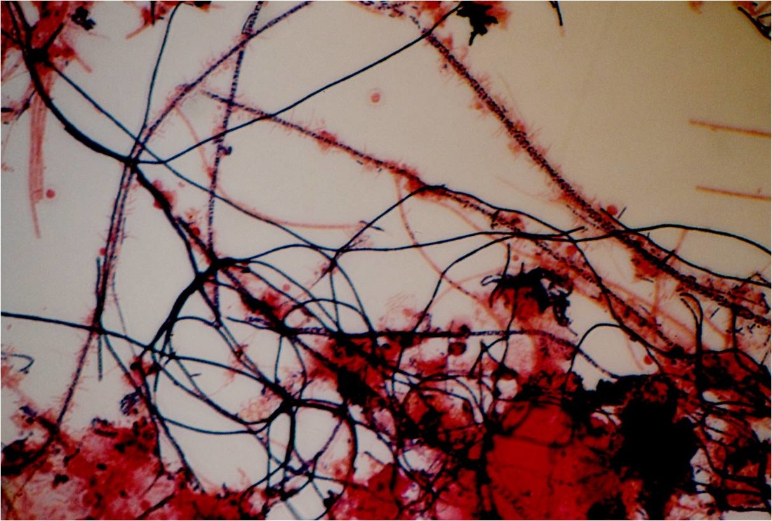
Neisser positive (purple) filament is Nostocoida limicola II. 980X
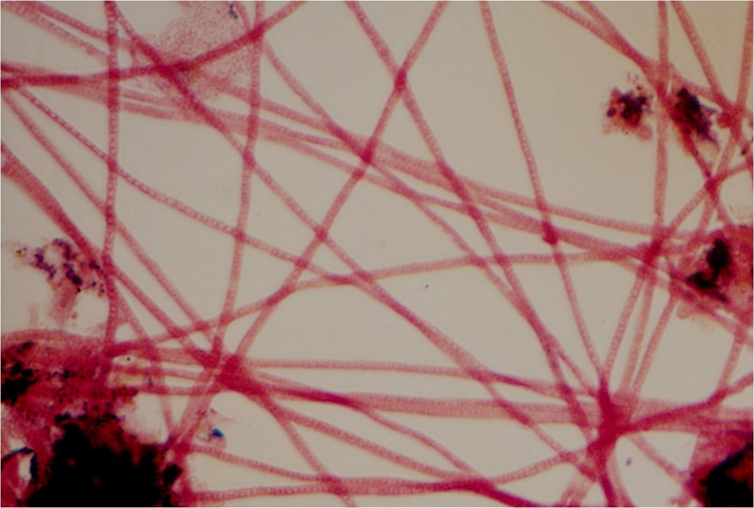
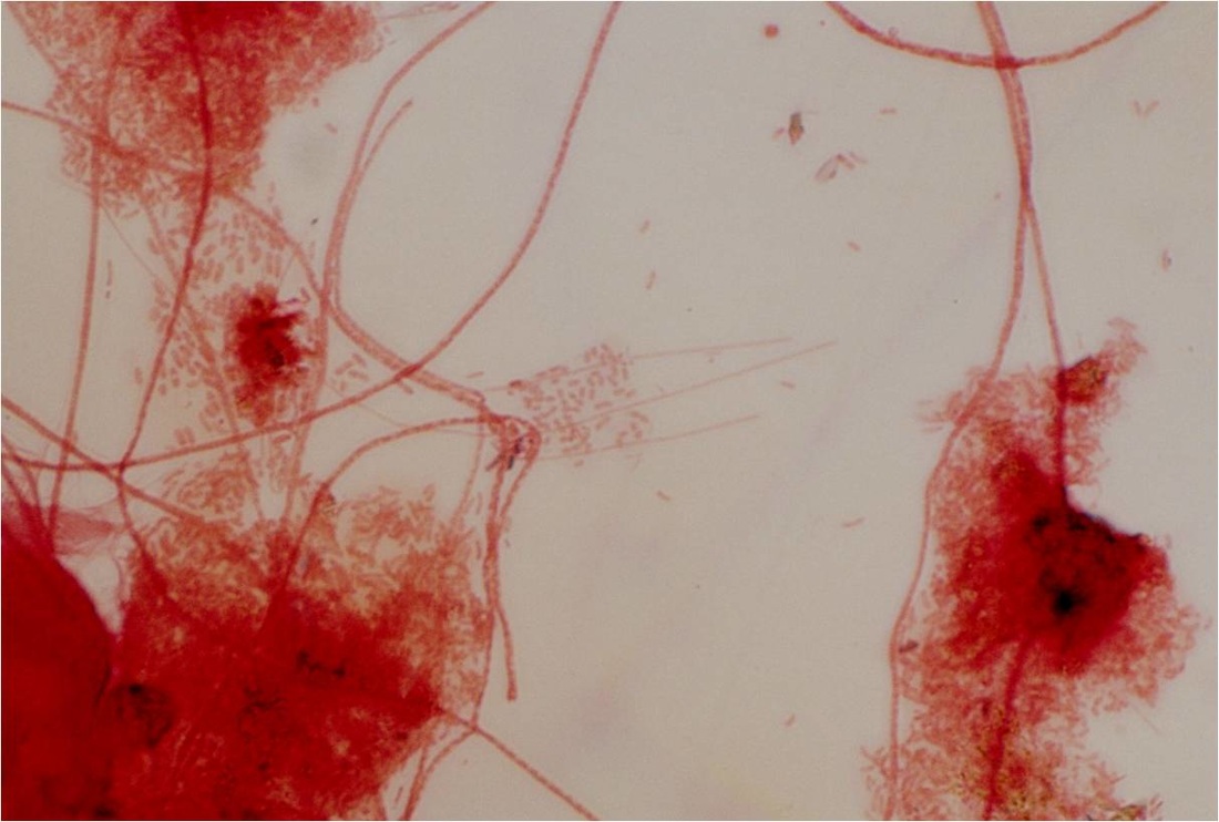
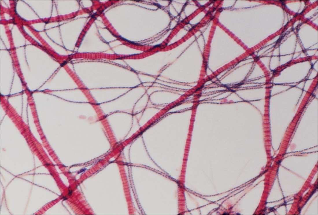
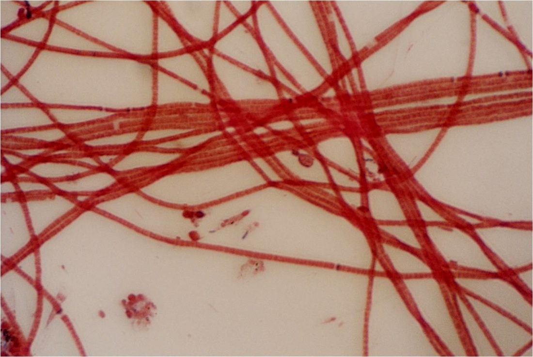
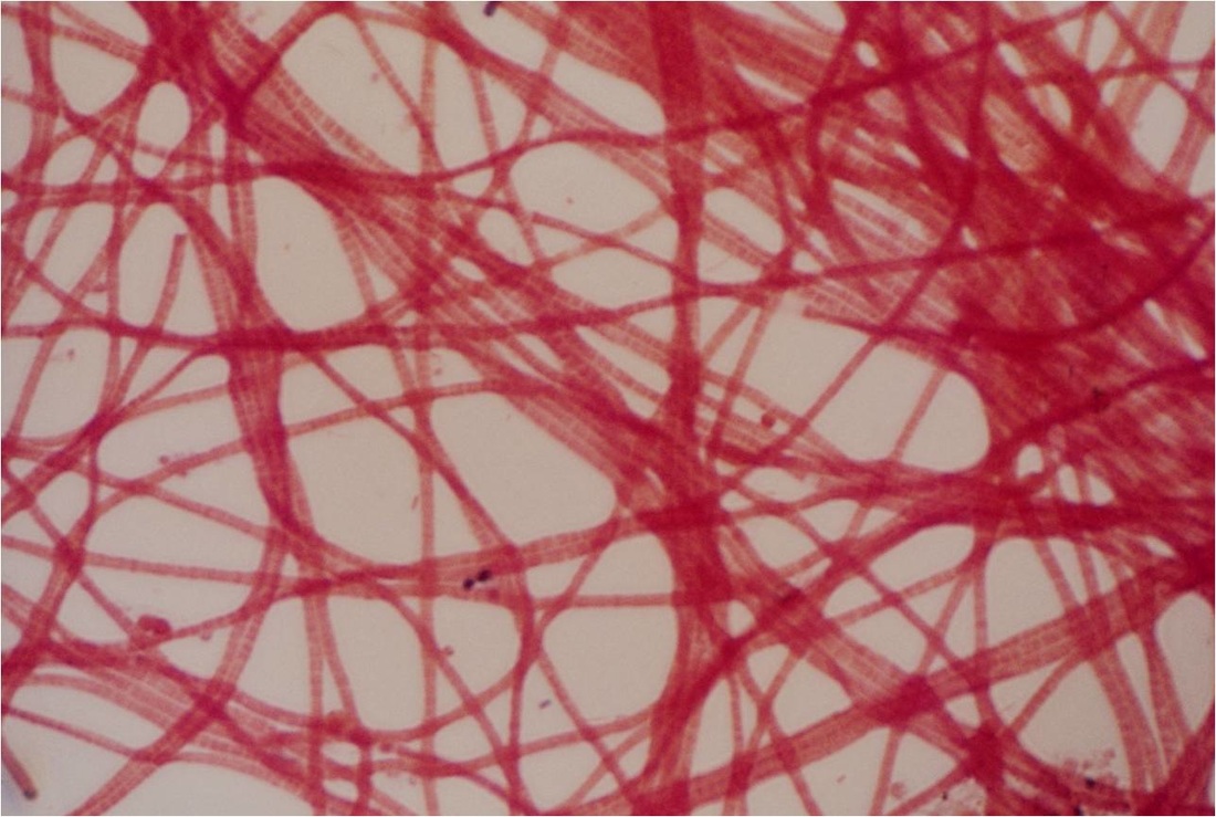
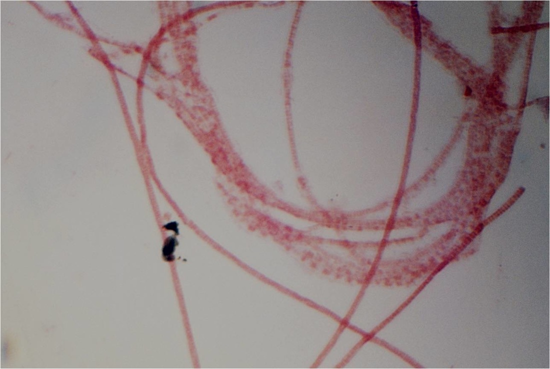
Gram-negative (red) filament O21N. 980X
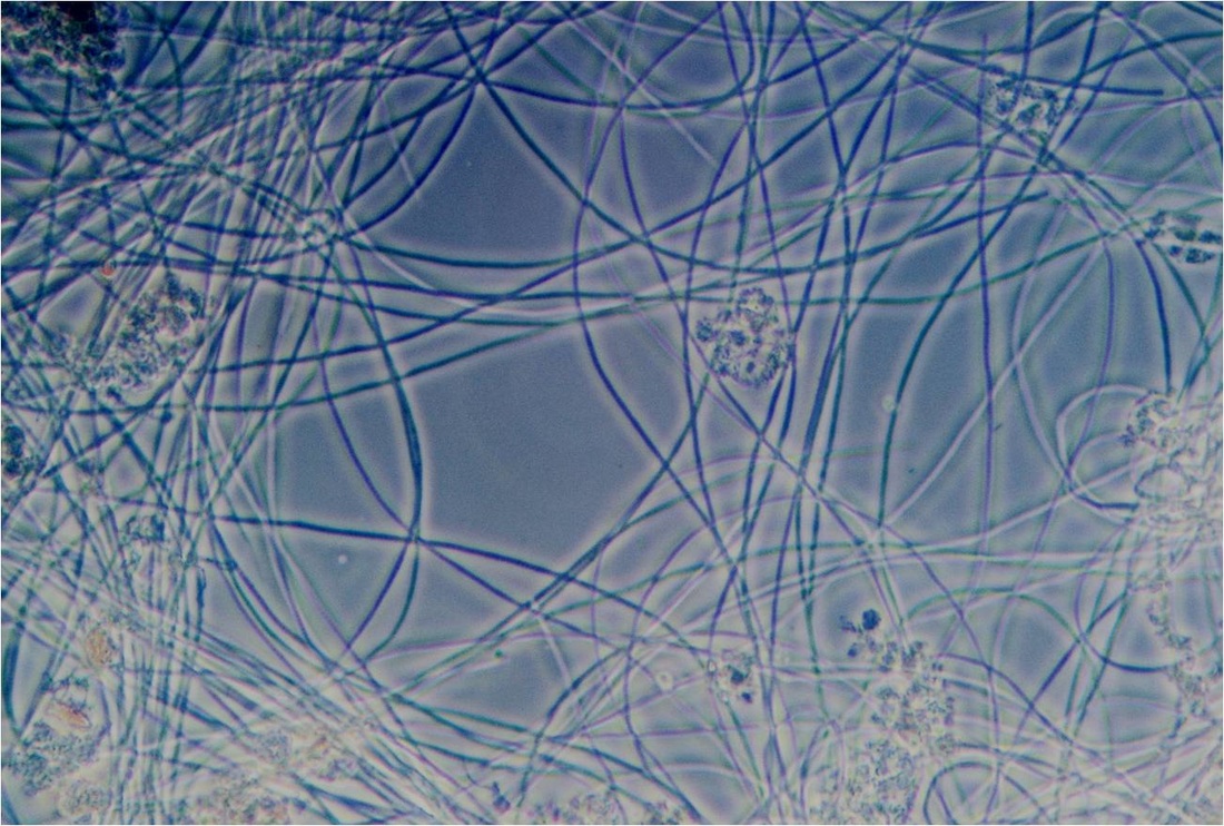
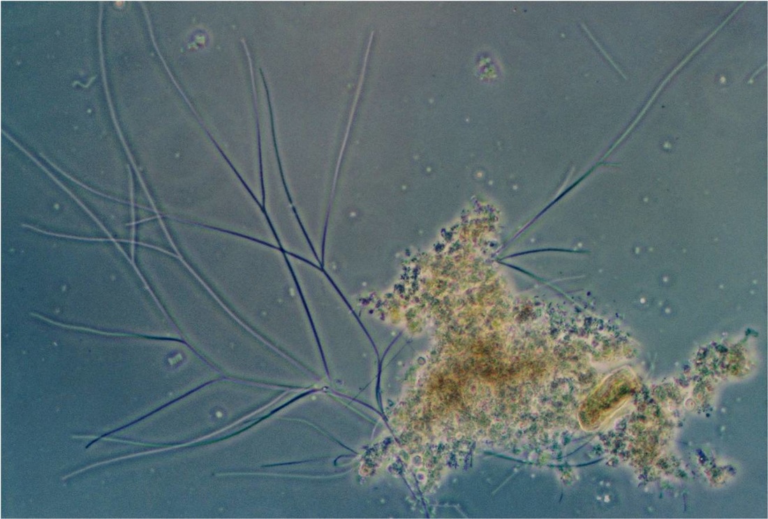
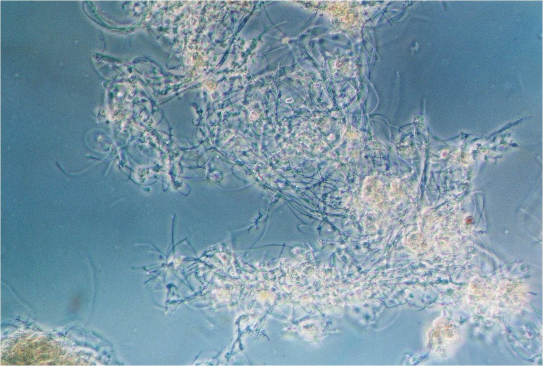
Low magnification wet-mount micrograph of filament O21N. 250X
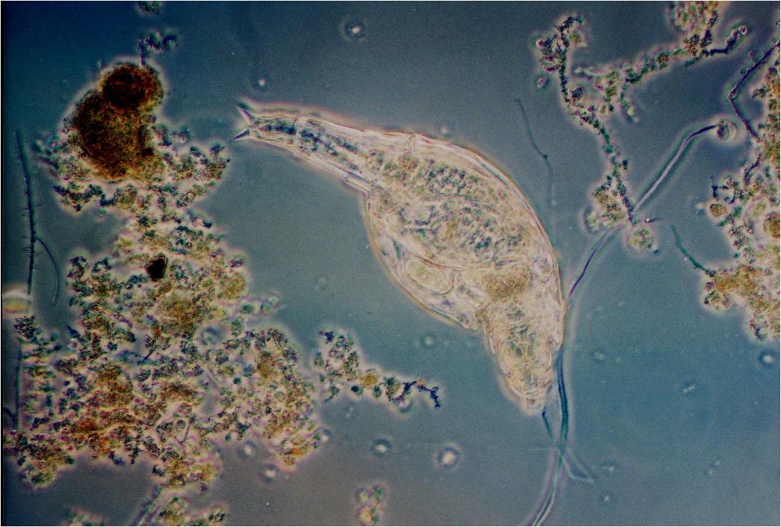
A rotifer in activated sludge. 250X
The falsely-branched filament Sphaerotilus natans. 250X
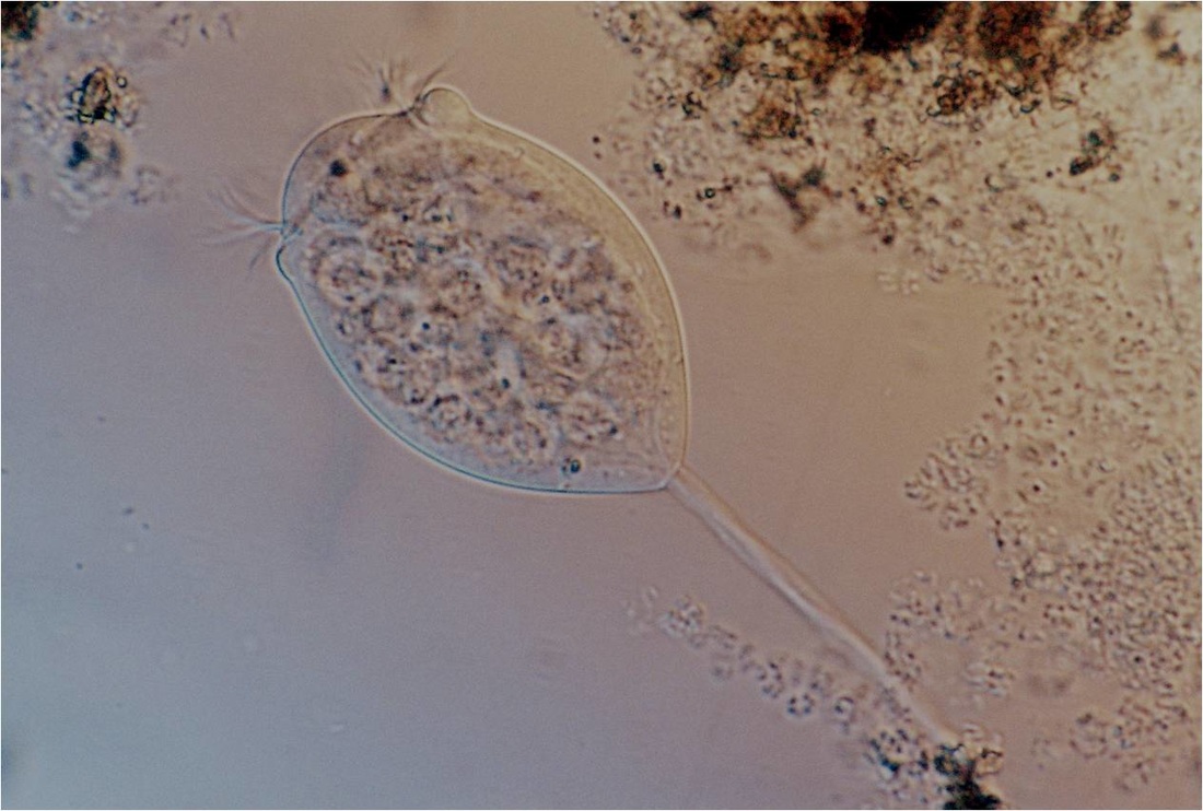
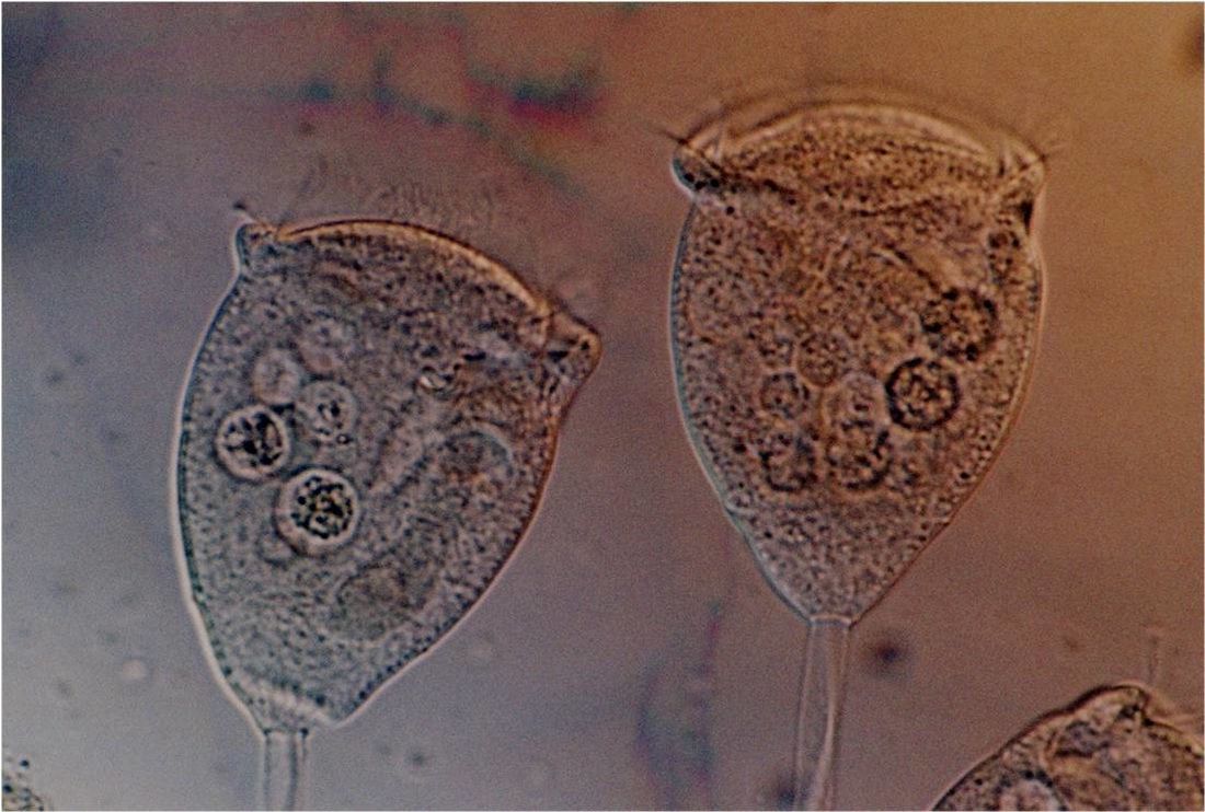
High magnification wet mount image of the common stalked ciliated protozoan, Vorticella. 640X
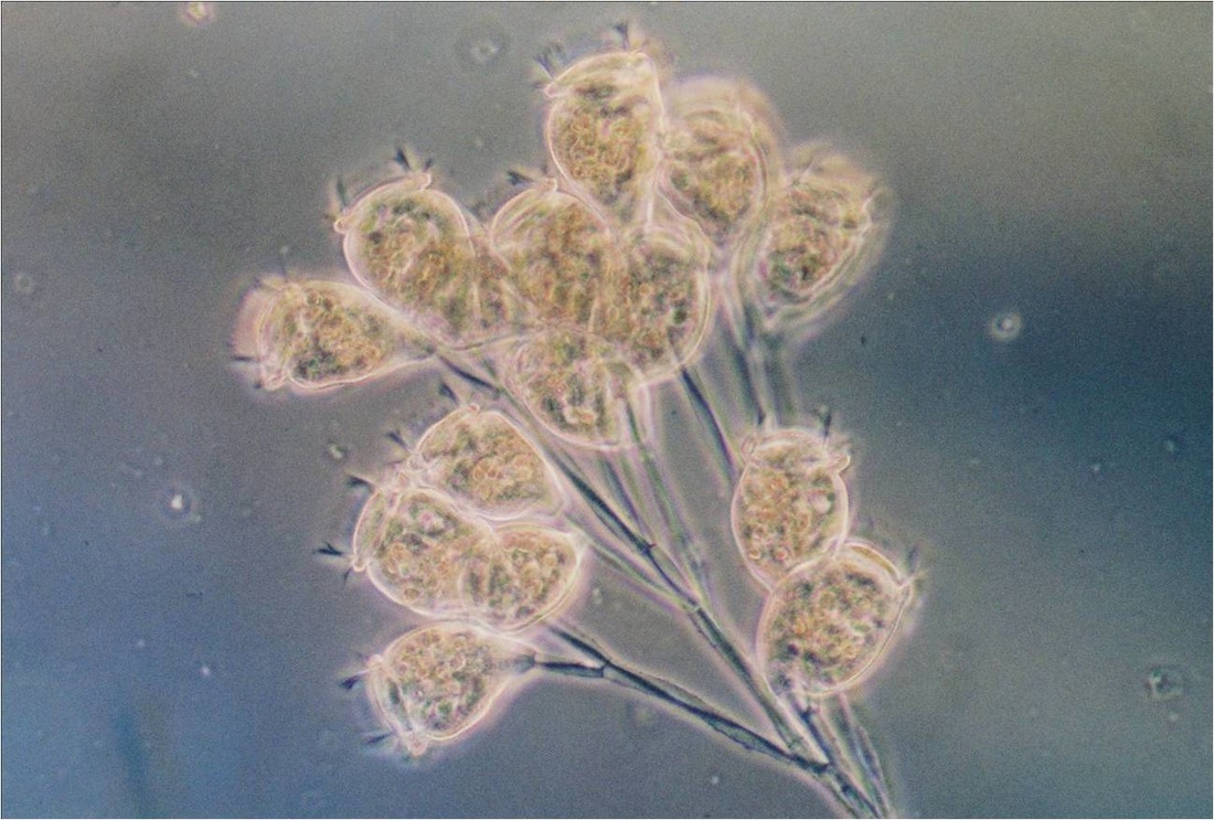
The colonial stalked ciliated protozoan Carchesium. 250X
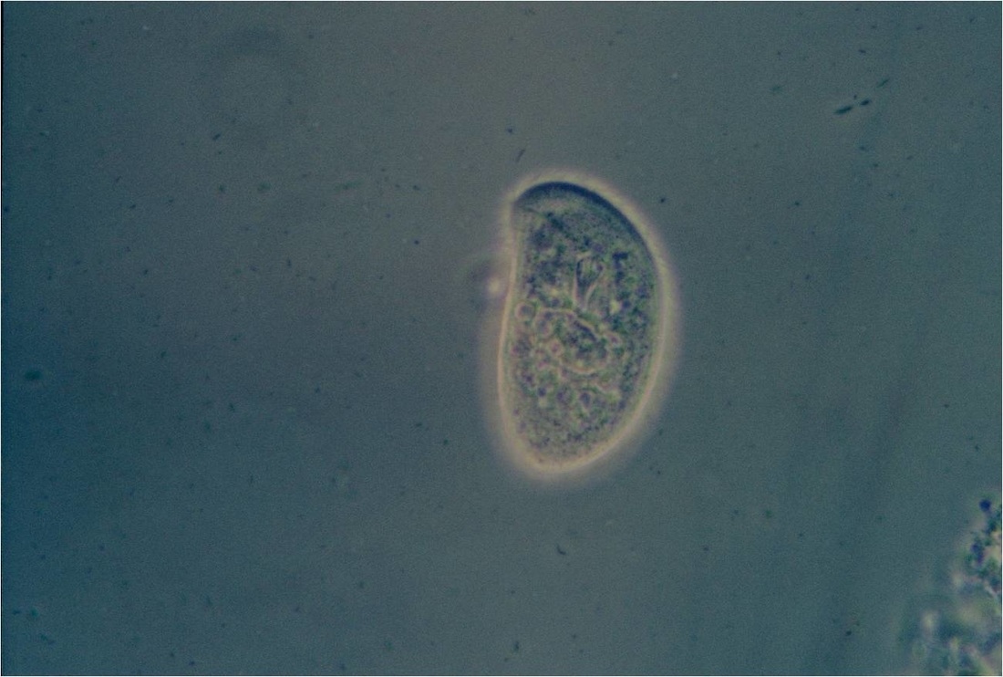
The free-swimming ciliated protozoan Chilodonella. 320X
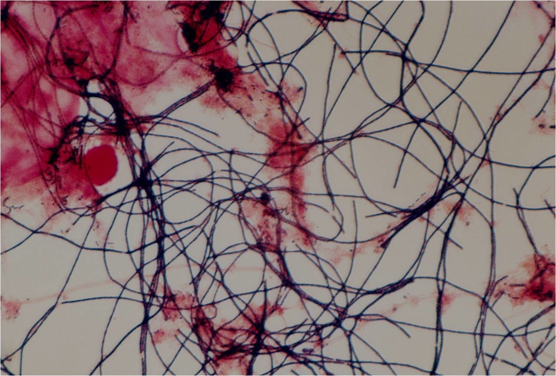
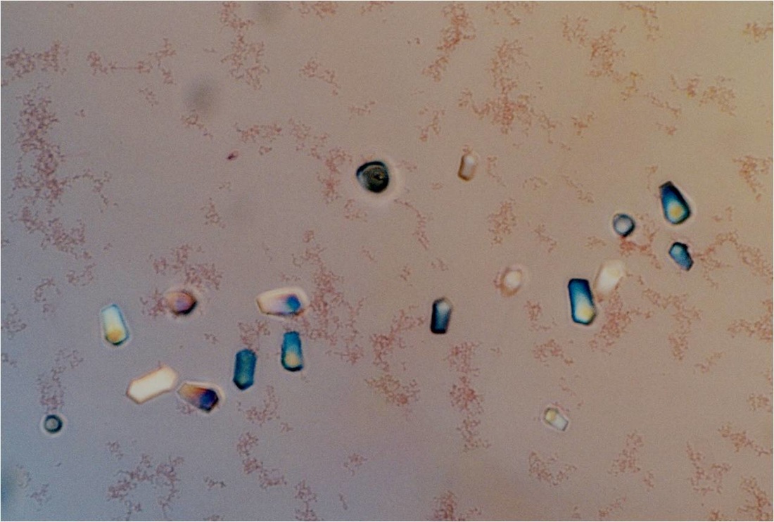
The Gram-positive (blue) filament is Microthrix parvicella. 980X
The very thin, horizontal, Gram-negative filaments are Haliscomenobacter hydrossis
Shown are two zooids (heads) of Vorticella at 640X
The thicker filaments with attached growth (see upper right) are type OO41. The thin blue filaments are Microthrix.
Very faint Gram-negative dispersed growth and inorganic particles (silt)
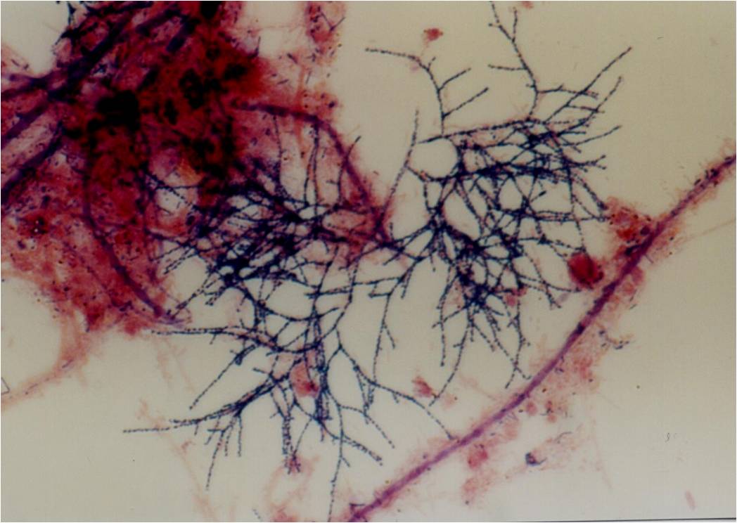
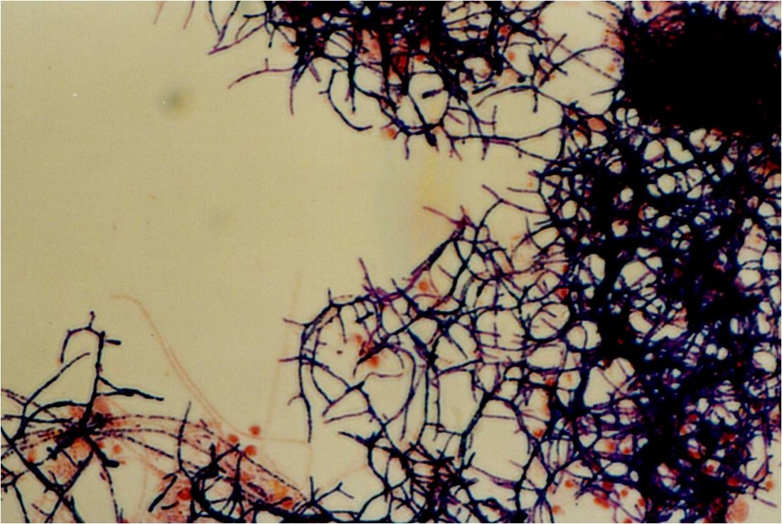
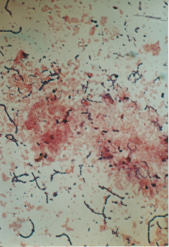
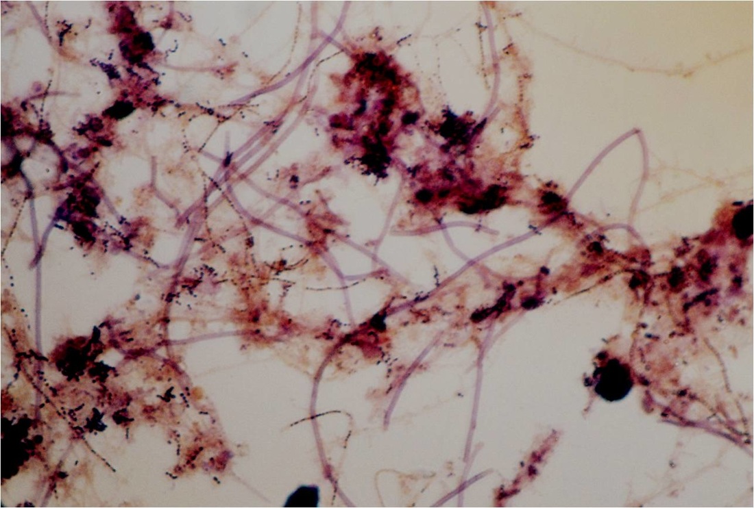
The Gram-positive (blue), branched filament is a nocardioform; most likely Nocardia or Gordonia
This micrograph shows how Microthrix can create flocs of low density, contributing to a bulking situation
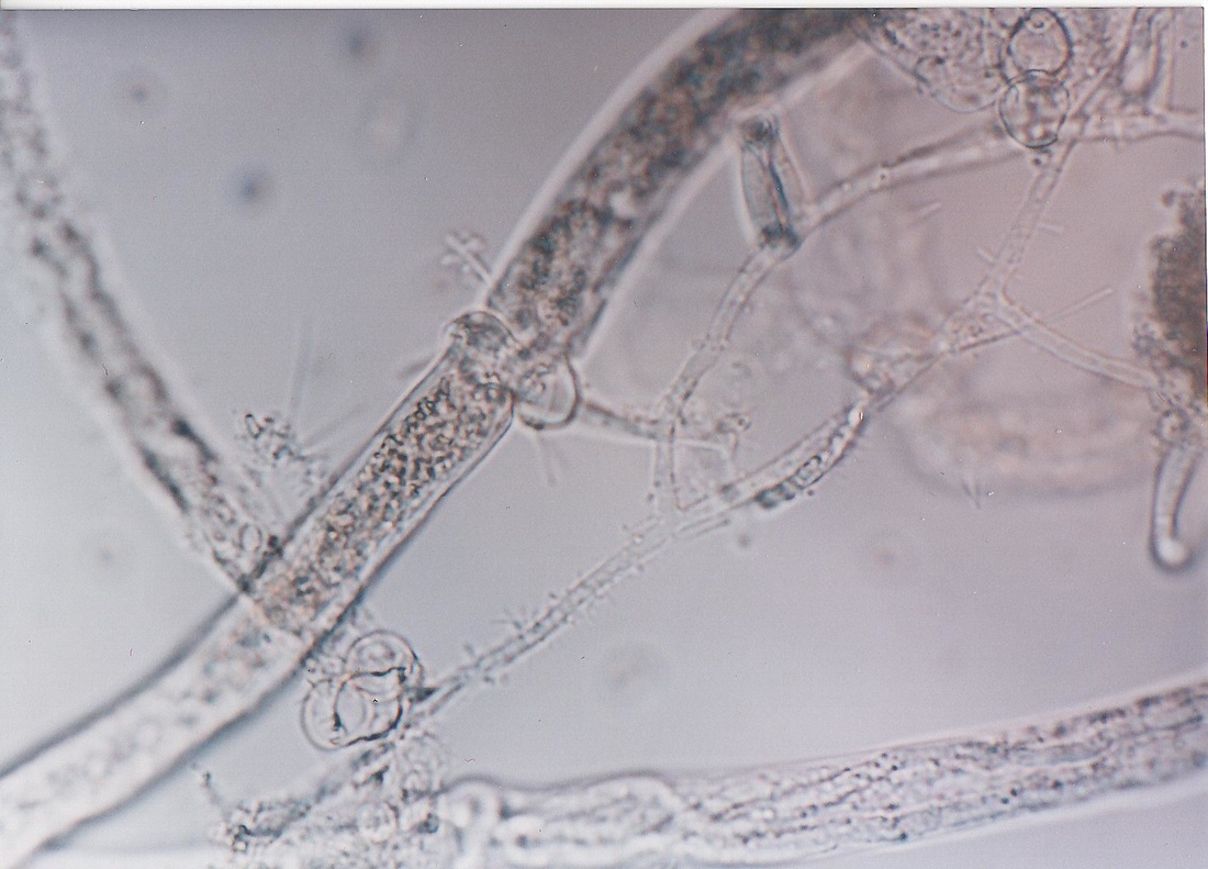
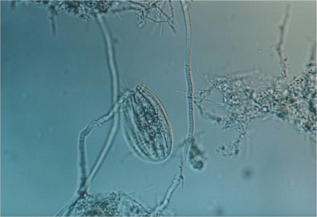
This cool image from an industrial facility shows a fungus that is predatory on nematodes.
The Gram-negative (red) filament is type O21N; the Gram-positive filament is Microthrix
This image shows the bacterial filament type O21N at a high level
The ovoid free-swimming ciliate in the center of the image is Euplotes
The nocardioform (most likely either Nocardia or Gordonia) in this image is present at a very high level. It is the blue, branched filament.
This image shows a very high concentration of healthy type O21N filaments.
Some of the O21N filaments in this image are coiled up; they are showing the effects of RAS chlorination.
Note the ragged looking O21N filaments on the left side of the image. This is a desired response to RAS chlorination.
This Gram stain shows loosely formed flocs and free bacteria; not the cohesive flocs that would indicate effective secondary treatment.
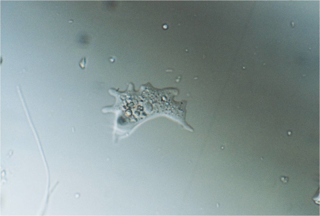
The organism in this low magnification wet mount is the naked amoeboid protozoan Mayorella
The purple (Neisser-positive) filament in this image is type 0092
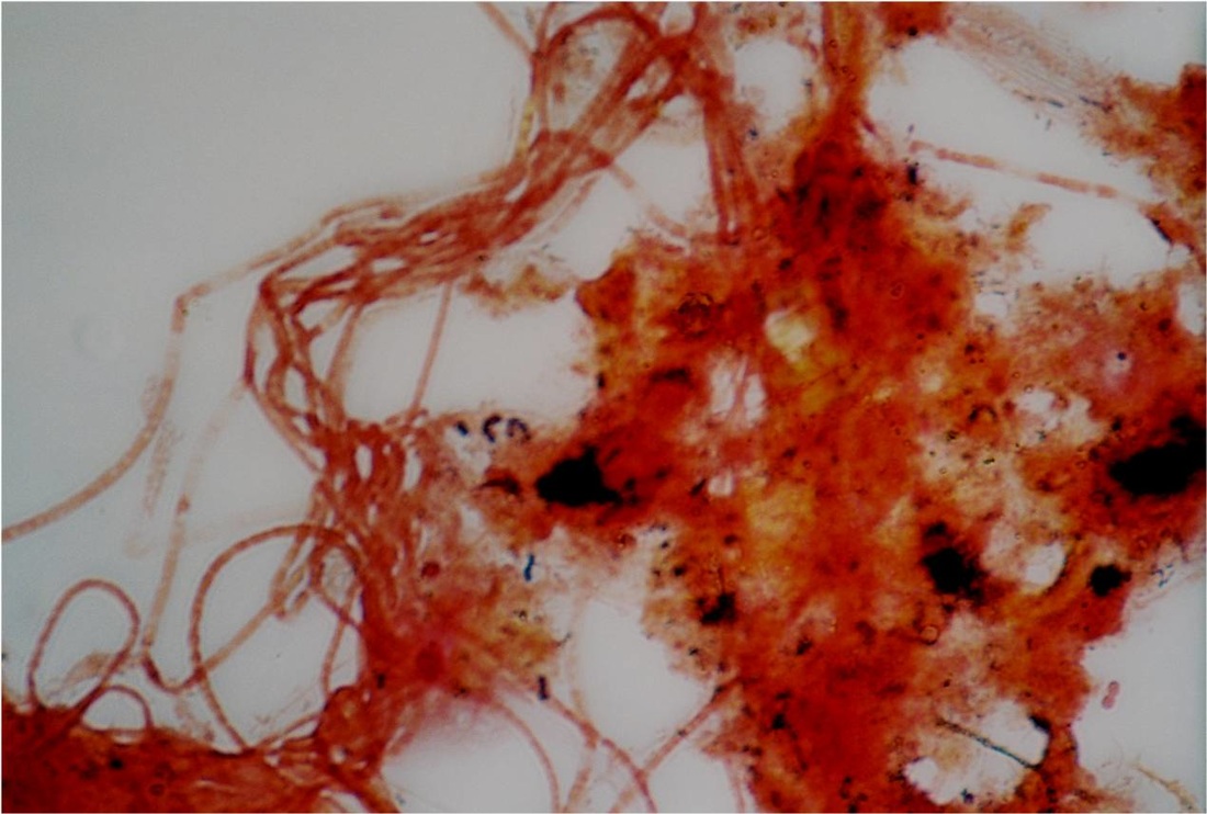
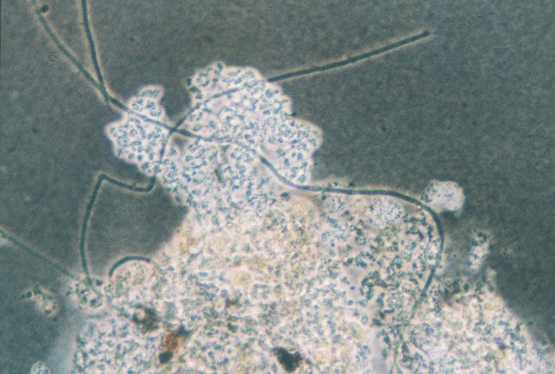
This is an India-ink wet mount. The bright area is a zoogloeal mass of bacteria with a considerable amount of exocellular polymeric substance present.